
Figure 4.
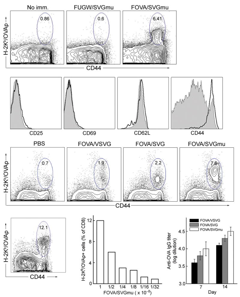
In vivo stimulation of antigen specific T cell and antibody responses in wild-type B6 mice following a subcutaneous injection of the DC-targeting lentivector FOVA/SVGmu. (a) B6 mice were immunized subcutaneously with 50×106 TU of either FOVA/SVGmu or FUGW/SVGmu (as a control). Mice without immunization (no imm.) were included as a negative control. Fourteen days post-immunization, spleen cells were harvested and analyzed for the presence of OVA-specific CD8+ T cells measured by H-2Kb-SIINFEKL-PE tetramer and CD44 staining. Indicated percentages are a percent of total CD8+ T cells. (b) Patterns of surface activation markers of OVA-specific CD8+ T cells (identified as tetramer positive cells) isolated from immunized mice 2 weeks post-injection were assessed by antibody staining for CD25, CD69, CD62L and CD44. Solid line, tetramer+CD8+ T cells from FOVA/SVGmu-immunized mice; shaded area, CD8+ T cells from non-immunized mice. (c) Naïve B6 mice were immunized by subcutaneous injection of 50×106 TU of the different lentivectors (FOVA/VSVG, FOVA/SVG, or FOVA/SVGmu). The injection of PBS was included as a control. Two weeks later, spleen cells were harvested and analyzed for the presence of OVA-specific CD8+ T cells measured by H-2Kb-SIINFEKL-PE tetramer and CD44 staining. Indicated percentages are a percent of total CD8+ T cells. (d-e) OVA-specific T cell responses seen in mice receiving different subcutaneous doses of FOVA/SVGmu. OVA-specific T cells were identified by tetramer staining as described in (a). (d) Percentage of OVA-specific CD8+ T cells following immunization with 100×106 TU of FOVA/SVGmu. (e) Dose responses of OVA-specific CD8+ T cells following injection of the indicated doses of FOVA/SVGmu. (f) OVA-specific serum IgG titer of B6 mice following immunization with 50×106 TU of the different lentivectors (FOVA/VSVG, FOVA/SVG, or FOVA/SVGmu). Sera were collected on day 7 and day 14 post-immunization and were analyzed for the titer of OVA-specific IgG using ELISA at serial 10× dilutions, starting at 1:100. The titer values were determined by the highest dilution at which the optical density was 2× standard derivations higher than that of the baseline serum at the equivalent dilution.