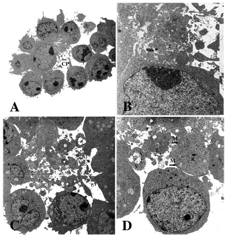
Figure 3.

Electron microscopy of CSVs. Intact clusters that had been incubated overnight without (A,B) or with (C,D) Ab 1F5. Cells were gently fixed with glutaraldehyde, to preserve the natural clusters, and then embedded in agarose in suspension and examined by electron microscopy. The space between the cells contains a heterogeneous mass of cytoplasmic vesicles, independent of the presence of Ab. A cytoplasmic projection (CP), shown in (A), suggests a possible origin of the vesicles. The cytoplasmic fragments contain a few organelles: mitochondria (M), lipid droplets (L), vacuoles (V) and strands of RER (R) are shown (D). Magnification: (A) × 7200; (B) × 31 200; (C) × 13 200; (D) × 21 000. (B) is rotated 90° relative to (A), and (D) is rotated 180° relative to (C).
