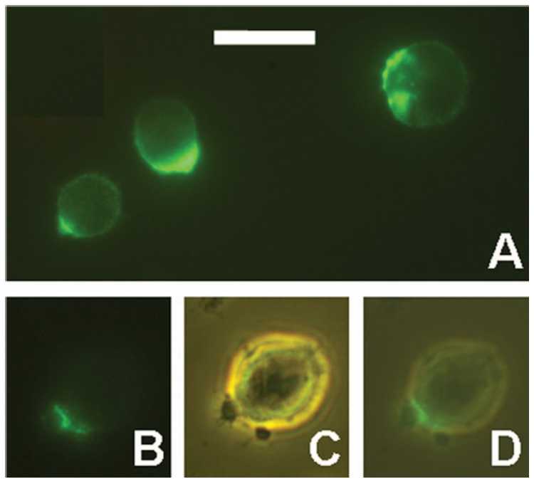
Figure 4.

Capping of CD20 and development of small CSVs. (A) RL cells were incubated for 3 h at 37°C with Alexa-488-1F5. Various staining patterns are shown. (B–D) RL cells were incubated for 1 h at 37°C with Alexa-488-1F5, washed, and maintained at room temperature for approximately 1 h until the photographs were taken. A cell with two small blebs is shown by fluorescence (B), phase contrast (C), and a merged image of the two (D). Scale bar = 20 µm.
