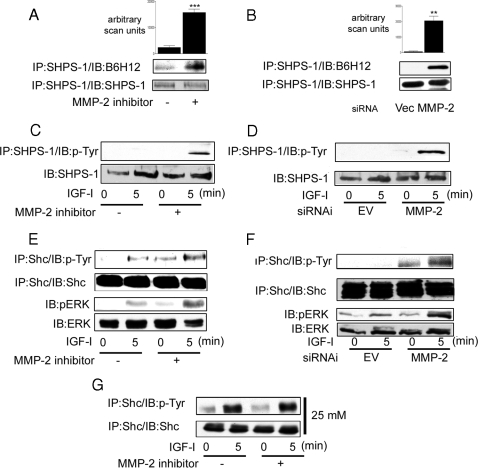
Figure 4.
Inhibition of MMP-2 Activity Enhances IGF-I-Mediated Signaling Events in SMC Grown in 5 mm Glucose
SMC grown to confluency in 5 mm glucose were incubated in SFM overnight. This was followed by a 4-h incubation in the presence of the MMP-2 inhibitor (3 μg/ml) before lysis and immunoprecipitation (IP). SMC expressing the siRNA-MMP-2 (MMP-2) or the empty vector control (Vec or EV) were grown to confluency in 5 mm glucose (or 25 mm glucose where stated). This was followed by overnight incubation in SFM followed by lysis and immunoprecipitation. Cells were treated with IGF-I (100 ng/ml) for the times indicated. A and B, SHPS-1 association with IAP was determined by immunoprecipitating cell lysates with an anti-SHPS-1 antibody and then immunoblotting with an anti-IAP monoclonal antibody B6H12 (top panel). Membranes were stripped and reprobed with an anti-SHPS-1 antibody (bottom panel). The graphs show the difference in IAP association with SHPS-1 expressed as arbitrary scanning units (mean ± sd, n = 3; ***, P < 0.05; **, P < 0.01). C and D, The extent of SHPS-1 phosphorylation was determined by immunoprecipitating with an anti-SHPS-1 antibody and then immunoblotting with an anti-phosphotyrosine antibody (p-Tyr) (top panel). Equivalent amounts of cell lysate were immunoprecipitated and immunoblotted with an anti-SHPS-1 antibody (bottom panel). E–G, The extent of Shc phosphorylation and activation of the MAPK pathway visualized by Western immunoblotting with antibodies that recognize the dual phosphorylated form of ERK1/2 was determined. In G, SMC were grown in 25 mm glucose.

