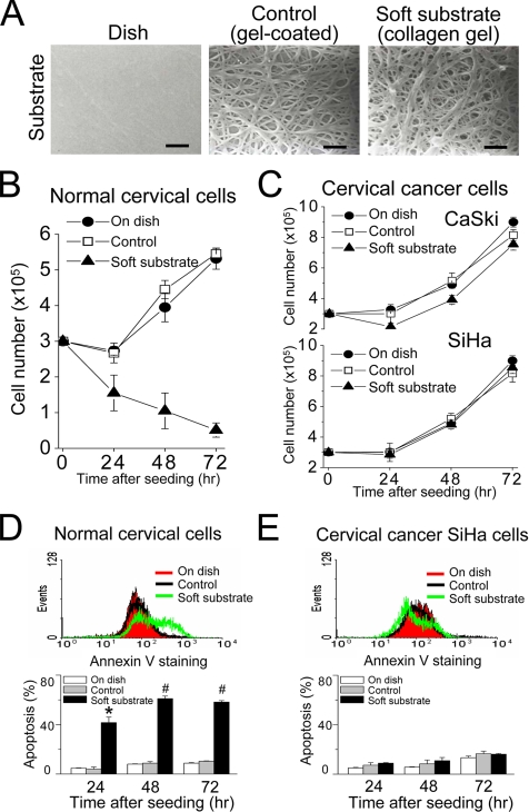
Figure 1.
Soft substrate regulates growth of normal cervical epithelial cells but not cervical cancer cells through apoptosis. (A) Cell culture system with collagen substrates of different elastic modulus. The culture dish was coated with a very thin layer of collagen gel (referred as “control group”) or overlaid with collagen gel (referred as “soft substrate”). Scale bars, 1 μm. (B and C) Different growth responses to soft substrate between normal cervical epithelial cells and cervical cancer cells (SiHa and CaSki). Each point represents mean ± SEM (n = 6). (D and E) Soft substrate induces apoptosis in normal cervical epithelial cells but not in cervical cancer SiHa cells. Top panels, annexin V staining of normal cervical epithelial cells and cervical cancer SiHa cells cultured on dish (On dish), collagen gel–coated dish (Control), or on collagen gel (On gel) for 4 h. X-axis indicates annexin V-FITC fluorescent intensity and Y-axis is the cell number. Bottom panels, apoptotic analysis was assessed by flow cytometry (FACScan) of propidium iodide (PI)-stained normal cervical epithelial cells and cervical cancer SiHa cells that were cultured on various conditions for 24, 48, and 72 h. Apoptosis indicates the cell population in sub-G1 phase of cell cycle. Each column represents mean ± SEM (n = 6). * p < 0.001; # p < 0.0001, compared with control group.

