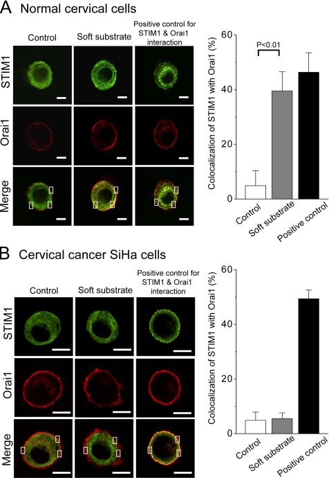
Figure 8.
The interaction between STIM1 and Orai1 detected by confocal images. Soft substrate stimulates the interaction between STIM1 and Orai1 in normal cervical epithelial cells (A) but not in cervical cancer SiHa cells (B). DNA plasmids of EGFP–STIM1 and mOrange–Orai1 were cotransfected into cells and then cultured on different substrate rigidities for 4 h. Soft substrate induced the aggregation and translocation of STIM1 toward the cell periphery to colocalize with Orai1 on the opposing plasma membrane of normal cervical epithelial cells. Cells treated with 2 μM thapsigargin were used as positive control for STIM1 and Orai1 interaction. Left panel, the representative confocal images. The inset indicates that a pixel-by-pixel colocalization analysis by FV-1000 software was used to assess levels of STIM1 colocalization with Orai1. The quantitative results are shown in the right panels. Scale bar, (A) 6 μm; (B) 10 μm.

