Figure 6.
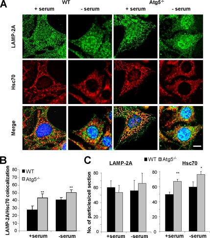
Changes in lysosomal supbopulations in Atg5−/− cells. (A) WT and ATG5−/− MEFs maintained in the presence or absence of serum for 16 h, as indicated, were methanol fixed (to eliminate soluble cytosolic proteins), blocked, and processed for double immunofluorescence with antibodies against LAMP-2A (green) and Hsc70 (red). Merged images of both channels are shown at the bottom. Bar, 5 μm. (B) Quantification of the fraction of LAMP-2A colocalizing with Hsc70 (lysosomes competent for CMA) in each condition. (C) Average number of puncta positive for LAMP-2A (left) and Hsc70 (right) per cell section in each condition. Values are mean of the quantification of 20 cells per condition in two independent experiments (*p < 0.05, **p < 0.001 compared with WT cells).

