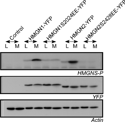
Figure 3.
Cell cycle-dependent phosphorylation of HMGN-YFP fusion proteins. HeLa cells were transfected with HMGN1-YFP, HMGN2-YFP, or the mutated fusion proteins HMGN1S2024E-YFP or HMGN2S2428E-YFP. Total cell extracts were prepared from log phase (L) or mitotic cells (M), and they were subjected to immunoblot analysis using antibodies recognizing HMGNs phosphorylated at the two serines located in their NBDs (Prymakowska-Bosak et al., 2001). Wild-type HMGN-YFP fusion proteins show mitotic specific serine phosphorylation in the NBD (top). Western analyses with actin demonstrate equal loading, and Western analyses for YFP (middle) demonstrates that all extracts contained comparable amounts of fusion proteins.

