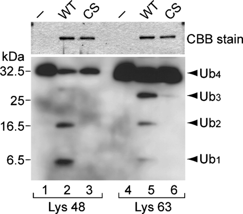
Figure 2.
DUB activity of USP30. In vitro deubiquitination assays of Lys-48– (lanes 1–3) and Lys-63–linked (lanes 4–6) Ub4 were performed in the absence (−; lanes 1 and 4) or presence of GST-USP30DUB (WT; lanes 2 and 5) or GST-USP30DUB-CS (CS; lanes 3 and 6). The reaction samples were analyzed by SDS-PAGE followed by Coomassie Brilliant Blue staining to detect the GST-fusion proteins used (top) and Western blotting with anti-Ub antibody (bottom). Arrowheads indicate the positions of tetra- (Ub4), tri- (Ub3), di- (Ub2), and mono-Ub (Ub1).

