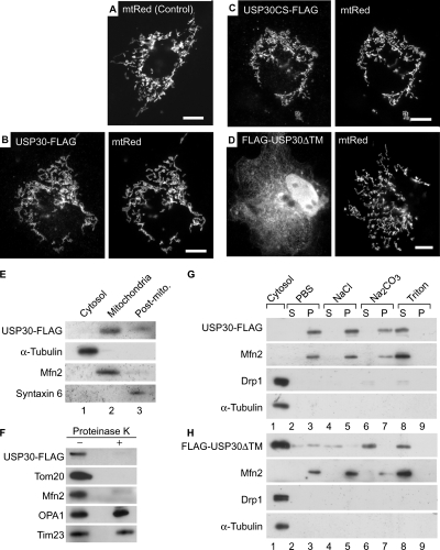
Figure 3.
Subcellular localization of USP30. (A–D) COS7 cells transiently transfected with matrix-targeted red fluorescent protein (mtRed) alone (Control; A) or together with USP30-FLAG (B), USP30CS-FLAG (C) or FLAG-USP30ΔTM (D) were stained with anti-FLAG antibody. Signals for anti-FLAG antibody (left) and mtRed (right) are shown. Bars, 10 μm. (E) The cytosolic, mitochondrial, and postmitochondrial (Post-mito.) fractions (5 μg of protein each) were analyzed by Western blotting with antibodies against FLAG, α-tubulin, Mfn2, and syntaxin 6. (F) The mitochondria-rich fractions of COS7 cells expressing USP30-FLAG were incubated with (+) or without (−) 100 μg/ml proteinase K and then used for Western blotting with antibodies against the indicated antigens. (G and H) Homogenates of COS7 cells expressing USP30-FLAG (G) or FLAG-USP30ΔTM (H) were fractionated into the cytosolic (lane 1) and membranous fractions. The membranes were incubated with PBS (lanes 2 and 3), 1 M NaCl (lanes 4 and 5), 0.1 M Na2CO3 (pH 11.5, lanes 6 and 7), or 1% Triton X-100 (lanes 8 and 9) followed by ultracentrifugation to separate soluble protein supernatants (S; lanes 2, 4, 6, and 8) from membranous pellets (P; lanes 3, 5, 7, and 9). The samples were analyzed by Western blotting with antibodies against FLAG, Mfn2 (a TM protein), Drp1 (a cytosolic protein), and α-tubulin (a cytosolic protein).

