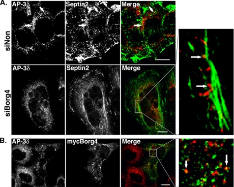
Figure 7.
Localization of AP-3δ in siBorg4 and myc-Borg4-treated cells. (A) HeLa cells were transfected with either control siNon or siBorg4, double labeled with antibodies against AP-3δ (red) and septin2 (green). (B) HeLa cells expressing myc-Borg4 were fixed, permeabilized, and labeled with antibodies against AP-3δ (red) and myc (green). Cells were then fixed and analyzed by fluorescence confocal microscopy. The arrows indicate AP-3δ–positive structures that overlap with septin2 (A) or myc-Borg4 (Figure 7B) are aligned along septin2-positive filaments. Bars, 10 μm.

