Figure 5.
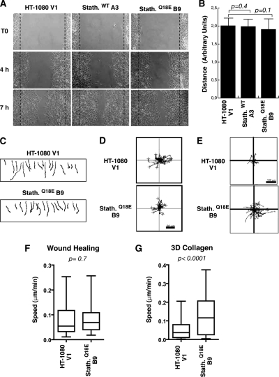
Stathmin expression exerts different effects on HT-1080 cells motility in 2D and 3D assays. (A) Typical images of HT-1080 V1, stathminWT A3, and stathminQ18E B9 clones during a wound-healing assay, in which the scratch was performed with a yellow tip. Bar, 100 μm. (B) Quantification of the migration distance covered by HT-1080 V1, stathminWT A3, and stathminQ18E B9 clones, over a 24-h period. (C–E) Orthotopically projected cell paths of HT-1080 cell clones, V1 (empty vector), and stathminQ18E clone B9, allowed to move toward a wound (C), randomly as sparse cells in 2D (D) or immersed in a 3D Coll I matrix (E), in serum-free medium. (F and G) Statistical evaluation (Mann-Whitney U test) of cell speed from experiments described in C and E. Median speeds were 0.07 and 0.09 μm/min for HT-1080 V1 and stathminQ18E B9, respectively, in the wound-healing assay (p = 0.7, Mann-Whitney U test) and 0.03 and 0.11 μm/min for HT-1080 V1 and stathminQ18E B9, respectively, in the 3D Coll I assay (p < 0.0001, Mann-Whitney U test).

