
Figure 1. TNF-α suppresses TGF-β1 induction in human dermal fibroblasts.
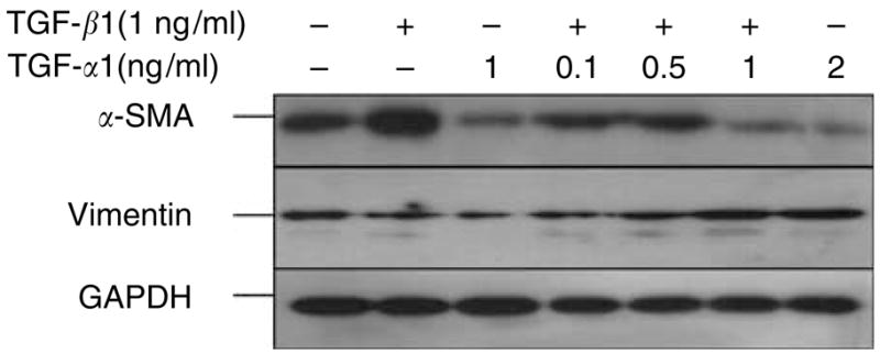
Normal human dermal fibroblasts (3 × 10⁵ cells/ml) were seeded in collagen I matrix (1 mg/ml). The fibroblast-populated collagen lattices (FPCLs) were treated with TGF-β1 (1 ng/ml), TNF-α (1 ng/ml), or combinations of TGF-β1 (1 ng/ml) and increasing concentrations of TNF-α for 96 hours under 0.1% serum conditions. Representative Western blots for α-SMA, vimentin, and GAPDH are shown.