
Figure 2. TNF-α suppresses TGF-β1 promotion of myofibroblast structural elements.
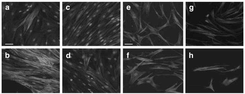
(a–d) Representative images of the immunohistochemistry staining for α-SMA. Normal human dermal fibroblasts (1 × 10⁵ cells) were seeded in collagen I matrix (1 mg/ml), which remained attach to the culture plate throughout the cytokine stimulation period. α-SMA was visualized using mouse monoclonal α-SMA FITC conjugated. (a) FPCL under control conditions at 96 hours. (b) FPCL treated with TGF-β1 (1 ng/ml) for 96 hours. (c) FPCL treated with TNF-α (1 ng/ml), and (d) FPCL treated with combination of TGF-β1 (1 ng/ml) and TNF-α (1 ng/ml). Bars = 50 μm (panels a–d). (e–h) Rhodamine-conjugated phalloidin staining for F-actin to examine the assembly of stress fibers. (e) FPCL under control conditions at 96 hours. (f) FPCL treated with TGF-β1 (1 ng/ml) for 96 hours. (g) FPCL treated with TNF-α (1 ng/ml), and (h) FPCL treated with a combination of TGF-β1 (1 ng/ml) plus TNF-α (1 ng/ml). Bars = 50 μm (panels e–h).