
Figure 6. TNF-α abolishes TGF-β1-induced myofibroblast contractility.
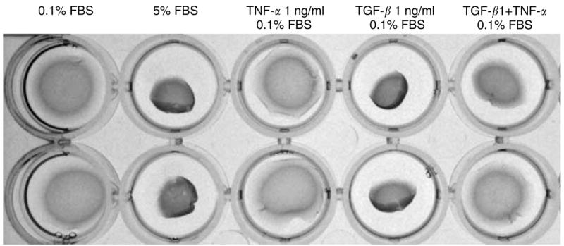
The stressed fibroblast-populated collagen lattice (FPCL) was prepared in a final cell density of 500,000 cells/ml plus collagen concentration of (1.5 mg/ml) and 0.6 ml volume per gel. The stressed FPCLs were treated under different conditions for 4 days. At the end of treatment, the stressed FPCLs were mechanically released from culture plate and allowed to contract rapidly within 1 hour. The pictures were taken at the end of contraction.