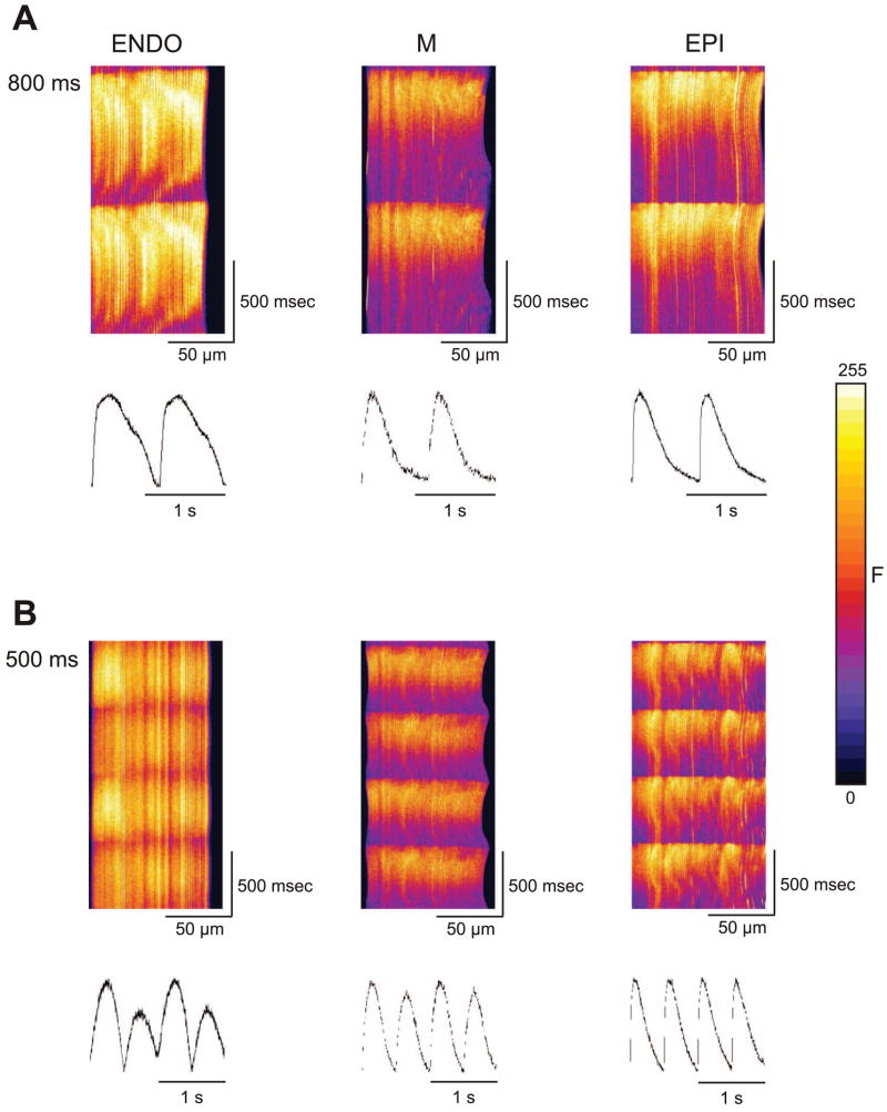
Fig. 4.
Representative line scans and time course of F/Fo recorded from the 3 ventricular cell types. A: scans at a cycle length of 800 ms. To induce alternans, cells were paced at progressively faster cycle lengths. B: line scans recorded at a cycle length of 500 ms show the appearance of alternans in the ENDO and MID region.

