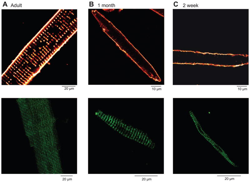
Fig. 6.
Ventricular cells isolated from dogs at various stages of development stained with the membrane dye di-8-ANEPPS (A–C, top). The t-tubular structure can be seen in adult cells (A, top). In contrast, ventricular cells from 2-wk-old dogs show no t-tubules (C, top), whereas 4-wk-old ventricular cells show t-tubules in a formative stage of development (B, top). Immunohistochemical localization of ryanodine receptors (RyR2) in ventricular cells (A–C, bottom) are shown. The adult ventricular cell shows prominent bands of RyR2 staining at the level of the t-tubules, suggesting a well-developed SR (A, bottom). The RyR2 organization in ventricular cells from 2-wk-old dogs (C, bottom) does not appear as well organized.

