
Figure 4.
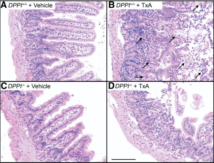
Deletion of DPPI inhibits TxA-induced enteritis. Images show the ileum. (A) DPPI+/+ vehicle control, (B) DPPI+/+ TxA, (C) DPPI−/− vehicle control, (D) DPPI−/− TxA. Note the normal villous structure in control groups. In DPPI+/+ mice, note loss of villi, infiltration of granulocytes, and severe necrosis (arrows), pathologic effects that were diminished in DPPI−/− mice. Scale bar = 100 μm.