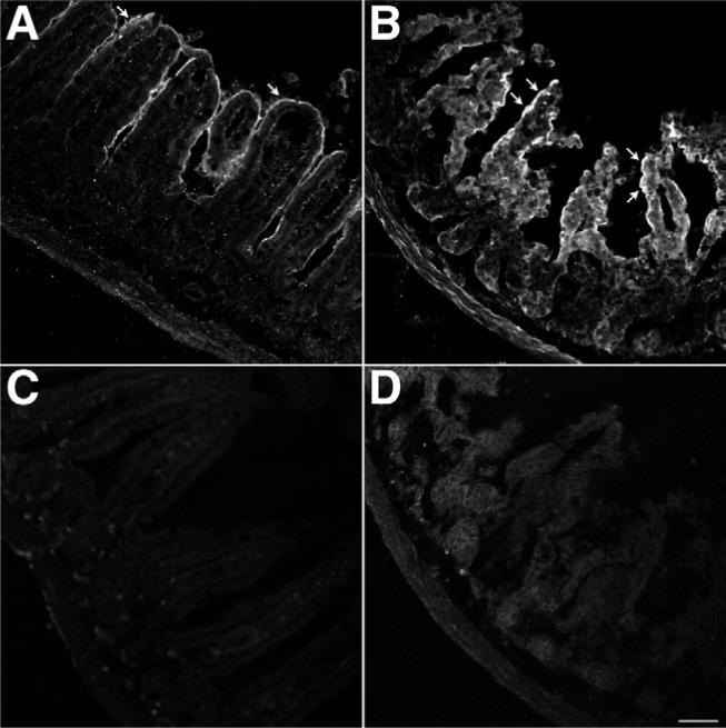
Figure 7.

Effects of TxA on PAR2 expression. Ileal loops formed in wild-type mice were injected with (A and C) vehicle or (B and D) 1.0 μg TxA. Tissue was stained for PAR2 using a mouse anti-PAR2 antibody 9717 and (A and B) a goat anti-rabbit secondary antibody conjugated to FITC or (C and D) the goat anti-rabbit secondary antibody conjugated to FITC alone. Images were acquired by confocal microscopy and are unmodified. The images shown in A and B were acquired under identical conditions to optimize the quantification of pixel intensity above baseline. The images shown in C and D were acquired under identical conditions to optimize tissue visualization. (A) Basal PAR2 expression with positive enterocytes (arrow). (B) TxA markedly up-regulated PAR2 expression in enterocytes (arrows). Omission of the mouse anti-PAR2 antibody 9717 eliminated specific staining in both (C) vehicle-treated ileum and (D) TxA-treated ileum. Scale bar = 50 μm.
