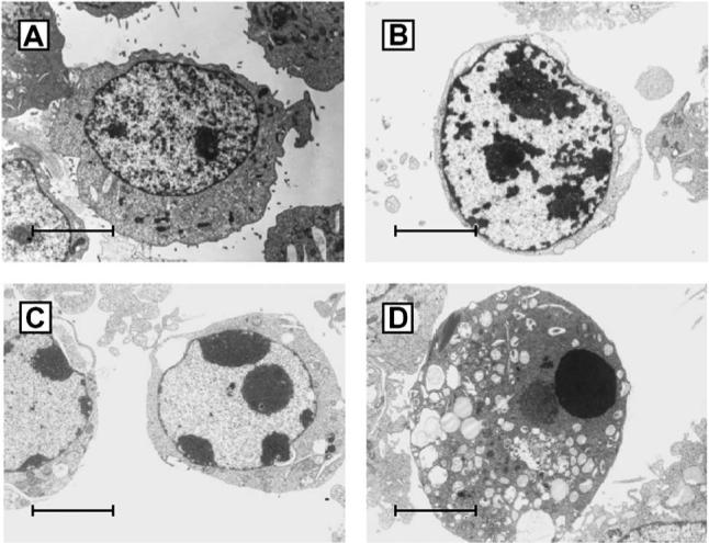
FIG. 1.

Electron microscopic appearance of uninfected (A) and apoptotic T3D-infected L929 fibroblasts (B–D). Note the progressive margination and compaction of the nuclear chromatin (B,C) and the eventual complete consensation of the nucleus (D). Despite profound changes in nuclear chromatin the cell membrane remains intact. From Tyler et al. (92) with permission.
