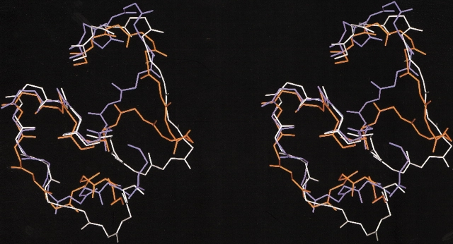
Figure 5.
Stereo figure of the superimposition of the main chains of the segment 153–176 in the structures of three P-I SVMPs that have an identical pattern of disulfide bonds but differ in their hemorrhagic potencies. BaP1, a weakly hemorrhagic SVMP (white); H2-proteinase, a SVMP devoid of hemorrhagic activity from the venom of Trimeresurus flavoviridis (blue); and acutolysin A, a SVMP with high hemorrhagic activity from the venom of Agkistrodon (Deinakgistrodon) acutus (orange).

