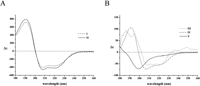
Figure 4.
Peptide C assumes α-helical structure upon binding to the small subunit of m-calpain. (A) The CD spectrum of the small subunit of m-calpain (21K) at 0.4 mg/mL was recorded in the absence (I) and presence (II) of an equimolar concentration of peptide C at 3 mM Ca2+ in water. (B) The difference spectrum obtained by subtracting the spectrum of the small subunit from that of peptide C + small subunit mixture (III). For a comparison, the spectra of peptide C in TFE (IV) and water (V), taken from Figure 1B ▶ and normalized to the given peptide concentration, are also shown.

