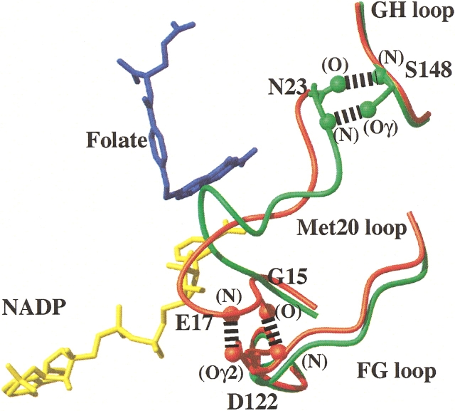
Figure 1.
The Met 20 loop conformations and hydrogen bonding patterns in the closed and occluded structures of E. coli DHFR. The figure shows a superposition of the α-carbon traces, for residues in the central regions of the Met 20, FG, and GH loops, of the E:folate:NADP+ (closed, shown in red) and E:folate (occluded, shown in green) complexes (Protein Data Bank accession numbers 1rx2 and 1rx7, respectively; Sawaya and Kraut 1997). The different hydrogen bonding interactions between the Met 20 loop and the FG and GH loops in the closed and occluded conformations are indicated. The location of bound folate (blue) and NADP+ (yellow) in the E:folate:NADP+ complex are shown.

