
Figure 2.
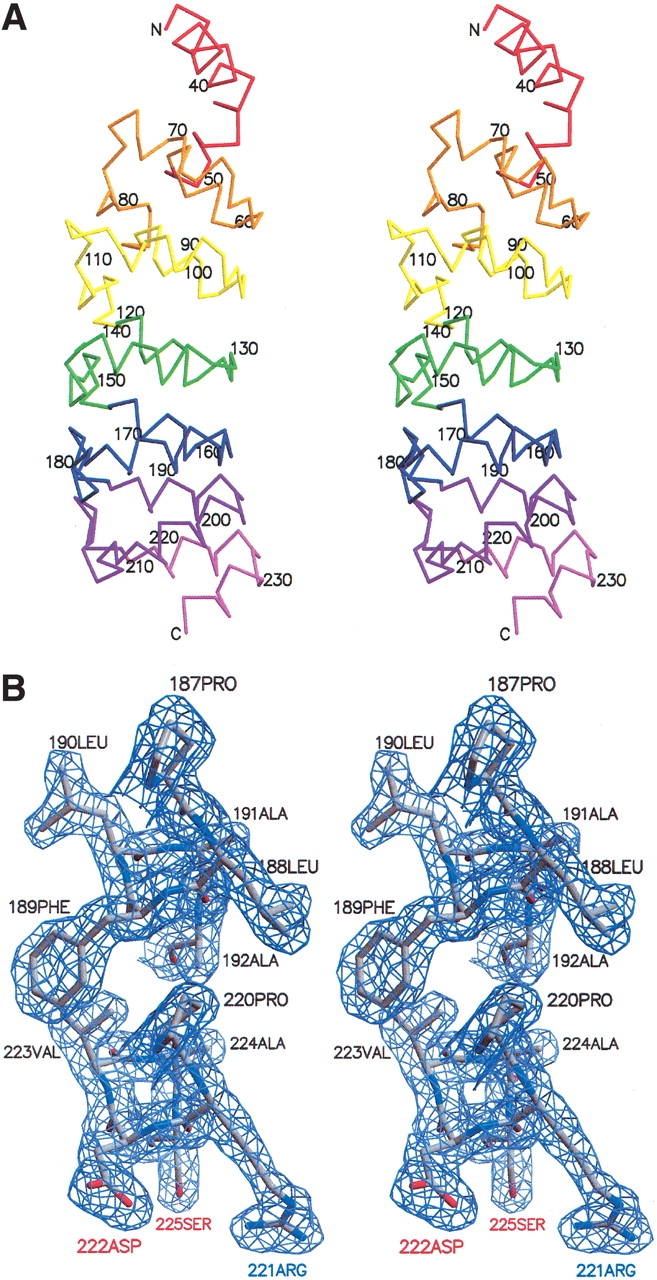
Crystal structure of the ankyrin repeat domain of the Drosophila Notch receptor. (A) Stereoview of the Cα trace of a single copy (chain A) of the ankyrin repeat domain. Individual ankyrin repeats are identified in different colors as in Figure 1 ▶. (B) Stereoview of representative portion of the electron density (2Fo-Fc) contoured at 1.2 sigma, showing the interface between the first helices of repeats 6 and 7. This figure was made using the programs MOLSCRIPT (Kraulis 1991), BOBSCRIPT (Esnouf 1997), and Raster3D (Merritt and Bacon 1997).