
Figure 7.
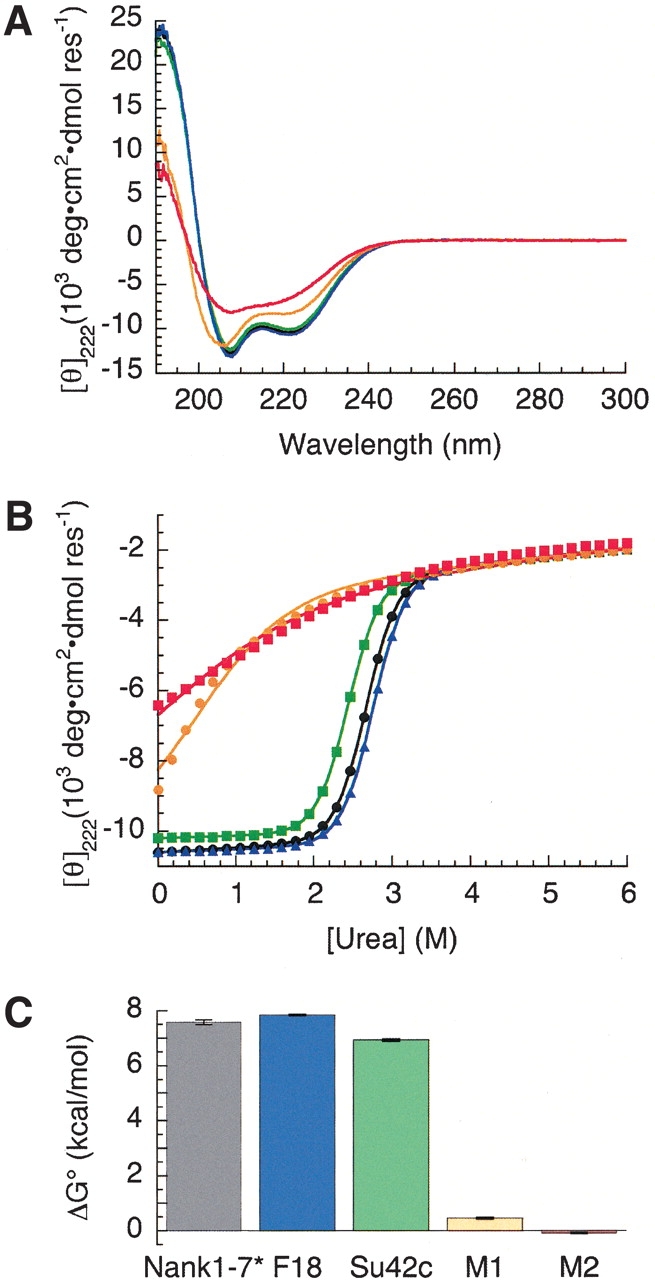
Structural stabilities of Notch variants. (A) Circular dichroism spectra of the Drosophila Notch ankyrin domain (black) and variants F18 (blue), Su42C (green), M1 (orange), and M2 (red). (B) Urea unfolding of the wild-type Drosophila Notch ankyrin domain (black circles) and variants F18 (blue triangles), Su42c (green squares), M1 (orange circles), and M2 (red squares). (C) Free energies of unfolding of the ankyrin domain variants. Colors are as in A.