
Figure 4.
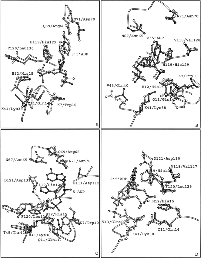
Superimposed structures of (A) the RNase A–3′,5′-ADP and EDN–3′,5′-ADP (PDB entry 1HI4) complexes; (B) the RNase A–2′,5′-ADP (molecule I) and EDN–2′,5′-ADP (PDB entry 1HI3) complexes; (C) the RNase A–5′-ADP and EDN–5′-ADP (PDB entry 1HI5) complexes, (D) the RNase A–2′,5′-ADP (molecule I) and ECP–2′,5′-ADP (PDB entry 1H1H) complexes. Residues and inhibitor molecules from the RNase A complexes are shown in gray and labeled using the one-letter code, while residues from EDN or ECP are shown in white and are labeled using the three-letter code.