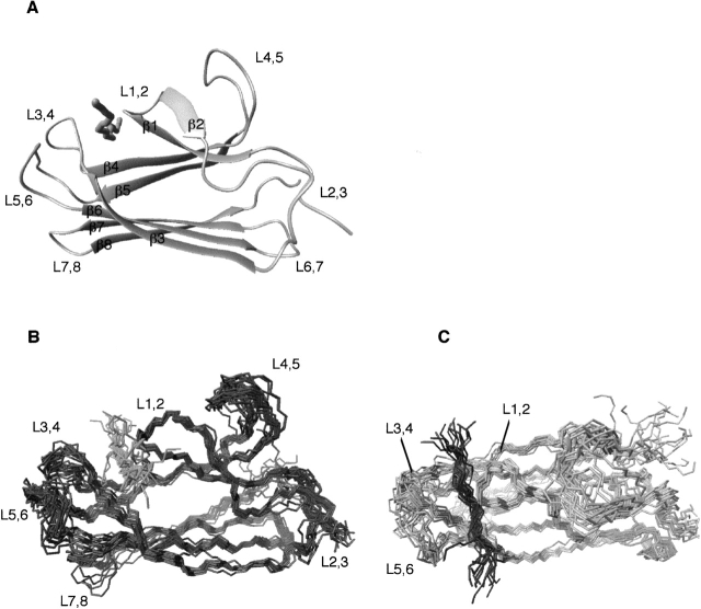
Figure 1.
(A) Ribbon diagram of the average structure of the DnaK β-domain (DnaK[383–507]) bound to the substrate peptide NRLLLTG. (B,C) Twenty energy-minimized conformers representing the solution structure of β-domain (DnaK393–507) bound to the peptide NRLLLTG are depicted. The conformers were superimposed using the secondary structure elements. The two views are rotated by 90°.

