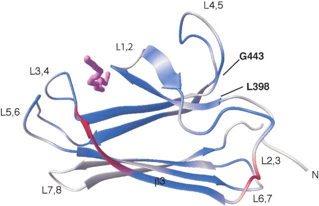
Figure 4.
Chemical shift differences observed upon titration of NRLLLTG form a path from loop L2,3 to the binding site. The residues in β3 that are unobserved due to exchange broadening (Gln 424–Ser 427) in the ligand-free state are colored red. The nuclei that exhibit chemical shift changes greater than or equal to 0.05 ppm for 1HN and/or greater than or equal to 0.30 ppm for 15N are colored blue. The peptide NRLLLTG is violet.

