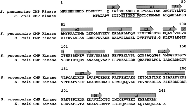
Figure 4.
Sequence alignment of the CMP kinases from S. pneumoniae and E. coli. The sequences are aligned using the three-dimensional structures of the proteins (Briozzo et al. 1998). The arrows and rectangles indicate the secondary structures of β-strands and α-helices, respectively. The dots in the sequence indicate gaps in the alignment. The rectangular open box indicates the phosphate-binding loop (P-loop).

