
Figure 3.
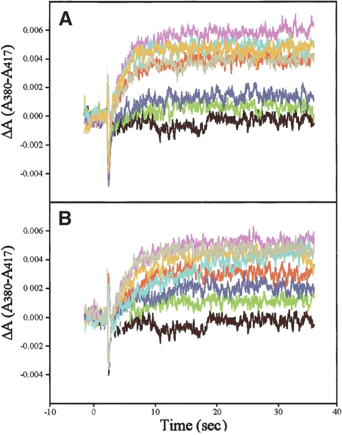
Extra-meta-II assays of flash-excited rhodopsin with antirhodopsin Fab antibodies with or without transducin-mimetic peptides. The difference in optical absorbance between the MII absorption maximum at 380 nm and at an isosbestic point between MI and MII at 417 nm (A380-A417), was measured versus time in hypotonically washed disk membrane suspensions. Formation of light-activated rhodopsin was triggered by a flash of 500 ± 20-nm light that bleached 12% of the rhodopsin. All samples contained 10 μM rhodopsin and 1 mM Gtα(340–350) wild-type peptide (IKENLKDCGLF; A), 10 μM Fab (if present). Black, rhodopsin baseline. Red indicates rhodopsin + peptide; green, rhodopsin + K42-41L and peptide; dark blue, rhodopsin and K60-46L and peptide; magenta, rhodopsin and K16-107C and peptide; light blue, rhodopsin and K16-111C + peptide; orange, rhodopsin and K16-155C and peptide; and grey, rhodopsin and 4B4 and peptide. (B) Shows 1 mM Gtγ(50–71) farnesyl peptide (EDPLVKGIPEDKNPFKELKGGC-farnesyl), 10 μM Fabs. The color key is the same as in A.