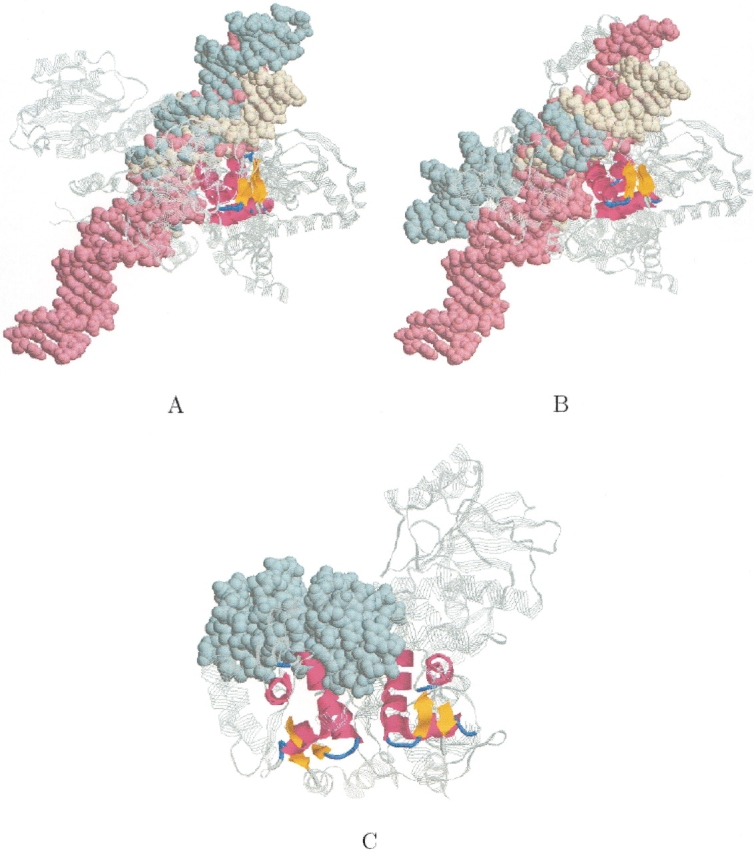
Figure 10.

Winged helix DNA binding domain. The figure shows the two different subset alignments that were obtained for the three winged helix DNA binding proteins (PDB: 1cgpA, 1fokA, 1ddnA) when we applied MASS to the DNA binding ensemble. The backbone of the proteins is colored gray. The cores of the alignments are colored by secondary structure. The DNA of PDB:1cgpA, 1fokA, and 1ddnA are colored in light yellow, light blue, and light pink, respectively. (A) The first detected subset alignment (also shown in Fig. 6E ▶). The DNAs of all the three complexes are well aligned. The core of the alignment is a winged-helix motif (three helices and a small β-sheet). (B) The second detected subset alignment. Only the DNAs of PDB:1cgpA and PDB:1ddnA are well aligned. The core of this alignment is also a winged-helix motif. (C) The figure shows that the two detected winged-helix motifs of PDB:1fokA are involved in DNA binding.
