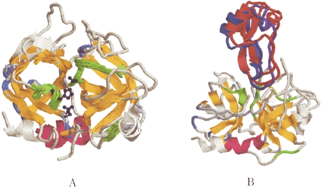
Figure 11.
Serine proteases. (A) The structural alignment of 10 serine proteinases. PDB:2pkaAB is shown completely in light yellow. The core of the alignment is colored by secondary structure and the three conserved loops are colored in green. The catalytic triad is also conserved (the triad of PDB:2pkaAB is depicted as ball and sticks and colored dark blue). Two of the conserved loops (55–59 and 189–197) are located in the active site. (B) The unbound docking, as obtained by PatchDock (Duhovny et al. 2002), between a serine protease kallikrein A (PDB:2pkaAB) and its bovine pancreatic trypsin inhibitor (PDB:6pti). The receptor PDB:2pkaAB is depicted as in A. The docked inhibitor, colored in red, is superimposed on the inhibitor of the crystal complex (PDB:2kaiI), colored blue.

