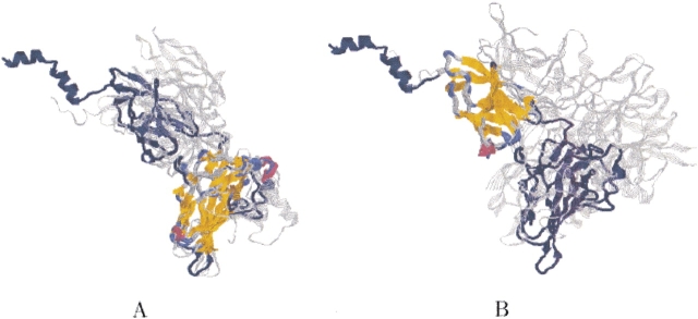
Figure 9.
Two-domains ensemble. The figure shows the two different structural conserved cores of the ensemble. The backbone of protein 1nfiA is shown in navy. The backbone of the other proteins is colored gray. The two structurally conserved cores detected by MASS are colored by secondary structure. (A) The first detected conserved core (part of the "p53-like transcription factors" domain). (B) The second detected conserved core (part of the "E set domains" domain).

