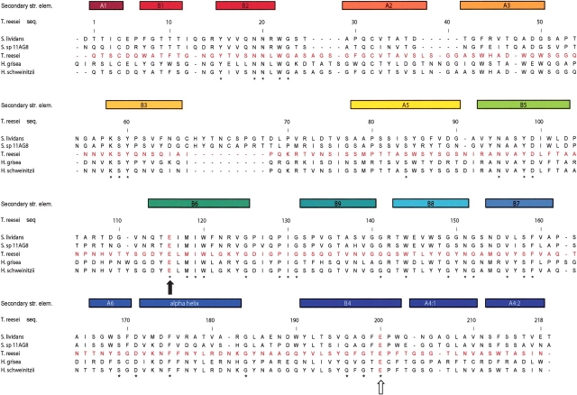
Figure 5.
Structure-based sequence alignment of five glycosyl hydrolase family 12 amino acid sequences with known protein structure. The secondary structure elements of the proteins, color-ramped from red at the N terminus to blue at the C terminus, are drawn at the top of the alignment. The position of the nucleophile and the acid–base in the sequences are indicated with filled and open arrows, respectively. The aligned protein sequences, with their GenBank or PDB access codes indicated in parentheses, are: Streptomyces lividans CelB2 (U04629, 2NLR); Streptomyces sp. 11AG8 Cel12A (AF233376, 1OA4); Humicola grisea Cel12A (AF435071, 1OLR); Trichoderma reesei Cel12A (AB003694, 1H8V); Hypocrea schweinitzii Cel12A (AF435068, 1OA3).

