Figure 6.


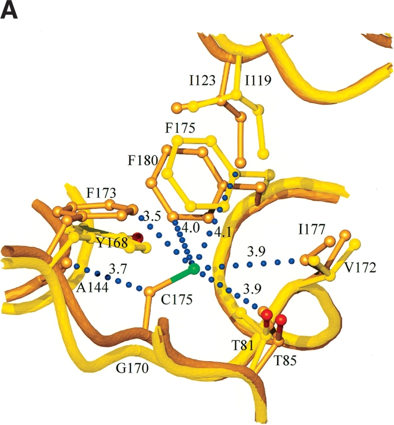
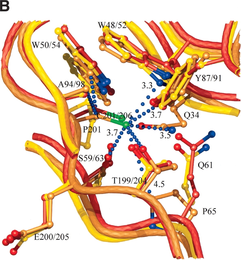
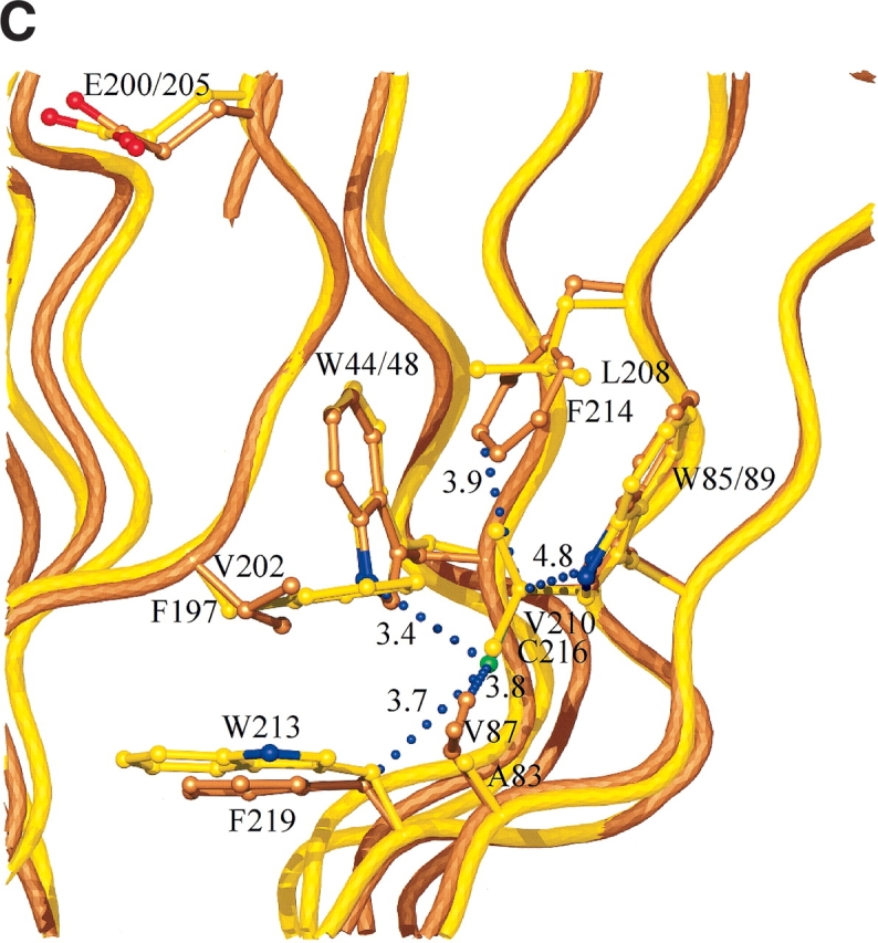
(A) Interactions and conformational changes close to residues 175 of the H. grisea Cel12A and residue 170 the T. reesei WT Cel12A structures. (B) Interactions and conformational changes close to residue 206 of the H. grisea Cel12A structure, and residue 201 of the T. reesei wild-type and P201C Cel12A structures. (C) Interactions and conformational changes close to residue 216 of the H. grisea Cel12A and 210 of the T. reesei wild-type Cel12A structures. The H. grisea Cel12A structures have carbon atoms colored goldenrod, and the T. reesei wild-type and P201C Cel12A structures have carbon atoms colored yellow and orange, respectively. The blue bubbles in A, B, and C indicate contacts to the free cysteine residues in H. grisea Cel12A.
