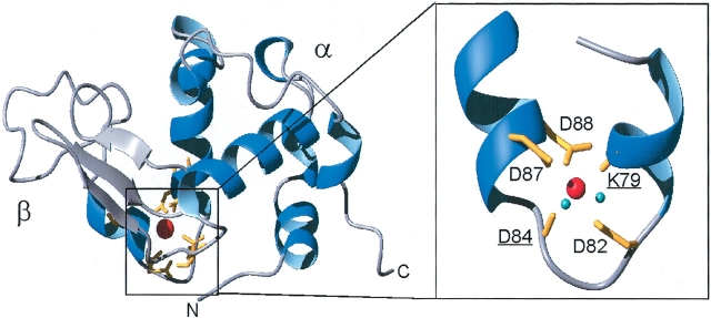
Figure 1.
3D structure of the α-lactalbumin Ca2+-binding site. Ribbon representation of the crystal structure of Ca2+-loaded human α-lactalbumin (Harata et al. 1999); PDB accession number 1B90. The Ca2+-binding site is magnified (rotated 180°) with the Ca2+-coordinating groups in yellow (K79 and D84 carbonyls, underlined, and D82, D87, and D88 carboxylate side chains). Ca2+ is red and Ca2+-coordinating water molecules are cyan. Figures were made in MOLMOL (Koradi et al. 1996) and rendered in Pov-Ray.

