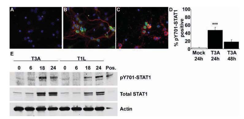
Figure 1.

Reovirus induces STAT1 phosphorylation at Y701 in primary neuronal cultures following infection. Cortical neuron cultures were mock or reovirus infected (MOI of 100). Dual-label immunocytochemical staining was performed to identify reovirus antigen σ3 (Texas Red; Virgin et al, 1991) and pY701-STAT1 (fluorescein) immunoreactive populations. Images represent σ3 and pY701-STAT1 immunoreactivity in mock (A) and T3A-infected neuronal cultures at 24 (B) and 48 (C) h post infection, at 400× original magnification. Very few neurons from mock-infected cultures provided any demonstration of pY701-STAT1 immunoreactivity. In T3A-infected cultures significantly higher numbers of pY701-STAT1 immunoreactive neurons were observed at 24 h post infection (B, D), although these numbers diminished by 48 h post infection (C, D). Western blot analysis of primary neuronal cultures infected with T3A and T1L demonstrated increased levels of pY701-STAT1 at 18 to 24 h post infection (E), accompanied by increases in total STAT1 protein levels at the same time points. Values represent the mean of four observations, with a minimum of 300 cells counted per observation, with vertical bars indicating the SEM. ***P < .001.
