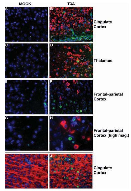
Figure 3.

Y701-phosphorylated STAT1 is expressed in areas surrounding brain injury following T3A infection. Two-day-old Swiss Webster mice were mock or T3A infected (i.c. inoculation of 1 × 10³ PFU) and sacrificed at 8 days post infection. pY701-STAT1 (fluorescein) and reovirus σ3 (Texas Red; Virgin et al, 1991) were identified by dual-label immunofluorescence (A-G). Fluorescence labeling was performed on brain tissue from mock(A, C, E, G) and T3A (B, D, F, H) infected animals. Images represent staining in the cingulate cortex (A, B), thalamus (C, D), and frontal parietal cortex (E, F at 400× and G, H at 630× [oil] original magnification). Arrow (D) indicates rare example of virus-infected cell demonstrating pY701-STAT1 immunoreactivity. Neuronal expression of activated STAT1 in T3A-infected animals was confirmed by dual label immunofluorescence of pY701-STAT1 (fluorescein) with the neuronal marker MAP-2 (Texas Red; I, J) in cingulate cortex of mock (I) and T3A- (J) infected mice.
