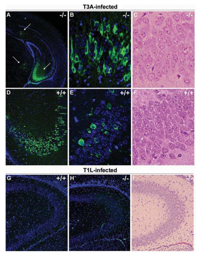
Figure 7.

Reovirus localizes to similar brain regions in STAT1 gene-deficient mice. Newborn STAT1 gene-deficient mouse pups and syngeneic controls (129SvEv) were T3A or T1L infected at 2 days of age with 100 PFU of virus. Brain injury was evaluated in H&E-stained sections and adjacent sections stained with monoclonal antibodies specific for σ3 (clone 4F2 for T3A-infected animals) or σ 1 (clone 5C6 for T1L-infected animals) with fluorescein-conjugated anti-mouse secondary antibody used to visualize labeling (A, B, D, E, F, G). Images represent H&E (C, F, I) and dual-label immunofluorescence (A, B, D, E, G, H) staining in the hippocampus of wild-type (+/+) and STAT1 gene knockout (-/-) mice at 25× (A), 100× (D, G-I), and 400× (B, C, E, F) original magnification. Viral antigen staining was not distributed differently between +/+ and -/- animals, although the frequency of immunoreactive populations was higher in -/- animals in hippocampal areas compared to +/+ groups. Viral antigen localization was predominantly neuronal in T3A-infected animals (B, D) and non-neuronal in T1L-infected mice (G).
