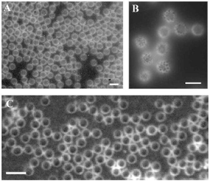
Figure 5.

A and B, Images of a population of lipid-coated monodisperse microbubbles incorporating DiI, illustrating the lipid and emulsifier phase coexistence. The scale bar represents 10 μm. C, Biotin-functionalized monodisperse microbubbles made without DiI, incorporating fluorescent avidin. Scale bar represents 5 μm.
