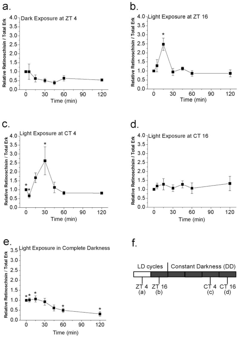
Figure 4.

Acute changes in illumination affect protein levels of retinoschisin. (a, b) E18 chick embryos in different preconditions were exposed to light or dark briefly for 0, 5, 15, 30, 45, 60, and 120 minutes. E18 embryos were under LD cycles for 7 days. (a) At ZT 4 (4 hours after lights on), brief exposure to darkness of various lengths of time did not affect the retinoschisin content. (b) At ZT 16 (4 hours after lights off), exposure to light for 15 minutes significantly increased the protein content of retinoschisin compared with other exposure time periods (0, 5, 30, 45, 60, 120 minutes). *P < 0.05. (c, d) E18 chick embryos, entrained under LD cycles for 5 days and kept in DD for 2 days, were exposed to the light on the second day of DD at CT 4 (c) or CT 16 (d) for 0, 5, 15, 30, 45, 60, and 120 minutes. (c) At CT 4 (subjective day), brief exposure to light for 30 minutes significantly increased retinoschisin compared with 0- and 5-minute light exposure (*P < 0.05). (d) At CT 16 (subjective night), acute light exposure did not change the retinoschisin content. (e) In E18 embryos that had been kept in complete darkness without any light experience, exposure to light for 60 or 120 minutes significantly decreased the protein expression of retinoschisin compared with 0-, 5-, and 15-minute light exposure (*P < 0.05). n = 5 to 6 for each time point. ANOVA with Tukey post hoc test was used to compare the retinoschisin protein level at different exposure time periods. (f) Different experimental paradigms from (a) to (d).
