
FIGURE 3.
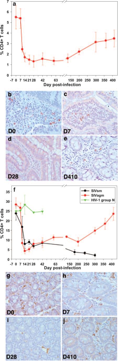
Longitudinal flow cytometric analysis of the percentage of intestinal CD4+ T cells in SIVagm-infected AGMs inoculated with SIVagm. A similar magnitude of CD4+ T cell depletion in the intestine (a) was shown as in pathogenic infection (f). Plots represent mean percentages of CD4+ T cells from animals in each group. Vertical lines with cross marks represent the SEM. Immunohistochemistry for CD4 confirms the loss of CD4+ cells in the gut by demonstrating the paucity of CD4+ T cells in lamina propria at day 7 p.i. (c), and day 28 p.i. (d), compared with either preinfection (b) or day 410 p.i. (e) when CD4+ T cells had recovered despite persistent viremia. Rh inoculated with SIVagm (red line) showed rapid and profound depletion of intestinal CD4+ T cells during acute infection (f) of the same order of magnitude as Rh infected with SIVsm (black line) (f). Controls inoculated with HIV-1 group N showed no change in CD4+ T cells (green line) (f). Vertical lines represent SEM. Immunohistochemistry for CD4 in macaques infected with SIVagm confirms the loss of CD4+ cells in the gut by demonstrating the paucity of CD4+ T cells in the lamina propria at day 7 p.i. (h) and 28 p.i. (i) compared with either preinfection (g) or day 410 (j), when CD4+ T cells recover. Original magnification, ×400.