FIGURE 2.
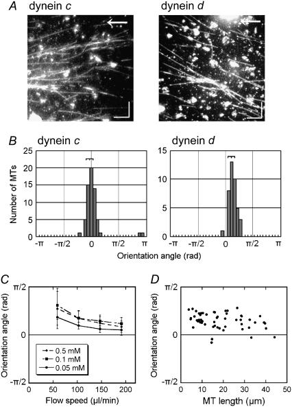
Effects of flow on the direction of microtubule translocation. (A) Dark-field images of microtubules on glass surfaces covered with dynein c or d, taken 3 min after application of shear flow (103 μl/min). Arrows indicate the direction of flow. (Bars) 10 μm. (B) Distribution of the angles between the direction of flow and the microtubule (orientation angle) at 3 min after initiation of the flow. The angle is defined as positive when the microtubule glides in a right-handed direction to the flow. The orientation angles were −0.1 ± 0.2 rad (n = 61) for dynein c and 0.4 ± 0.2 rad (n = 40) for dynein d. (C) Relation between the orientation angle (± SD) and the flow speed in the presence of various concentrations of ATP (0.05–0.5 mM) and 0.1 mM ADP in dynein d-translocated microtubules. (D) Dependence of the orientation angle on microtubule length in dynein d-translocated microtubules. Flow speed, 103 μl/min. The medium contained 0.5 mM ATP and 0.1 mM ADP in A, B, and D.

