
Figure 2.
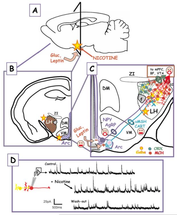
CNS sites of glucose, leptin, and (possibly) nicotine effects—up close and personal. (A) Glucose (Gluc), leptin, and nicotine signaling converge on the arcuate nucleus within the hypothalamus. The triangle shows the region from which the coronal sections in (B) and (C) are taken. (B) Higher magnification, coronal section showing the spatial relationship of the lateral hypothalamus (LH), the dorsal medial, and ventromedial hypothalamus (DM, VM) and the arcuate nuclus (Arc). ZI—zona inserta. (C) Glucose and leptin responsive neurons in the arcuate project to the LH. The first group of neurons, which are inhibited by the anorexigenic leptin signal (face with closed mouth), express the orexigenic peptides NPY and AgRP (face with open mouth). Leptin also excites a second group of neurons that contain the anorexigenic peptides, αMSH and CART. Both classes of Arc neurons project to the LH interacting with neurons and interneurons that express GABA (yellow), orexin (green), or MCH (red). Orexin and MCH neurons are orexigenic and project to other brain regions such as the mPFC, BF, VTA, etc. Key relays implicated in the regulation of feeding within the hypothalamic nuclei. The yellow stars represent possible loci of nicotine effects. (D) Nicotine treatment of LH neurons in vitro enhances neurotransmitter (GABA) release. The nicotine target is believed to be preynaptic nAChRs on GABAergic interneurons. The figure was modified from Jo and Role, 2002b, in press. (NPY, neuropeptide Y; AgRP, agouti-related peptide; αMSH, α-melanocyte-stiimulating hormone; CART, cocaine- and amphetamine-regulated transcript; ORX, orexin, MCH, melanin-concentrating hormone; f, fornix).