
Figure 3.
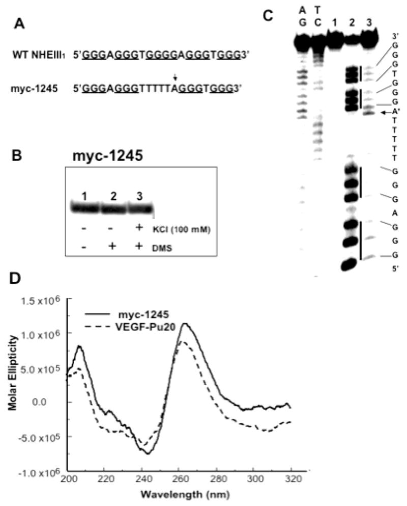
EMSA, DMS footprinting and CD spectrum of the sequence myc-1245. (A) Sequence of the wild type (WT) and mutant (myc-1245) NHEIII1 in the C-MYC promoter. (B) EMSA of the sequence myc-1245 pre-incubated under the conditions specified in the figure using a 16% native polyacrylamide gel. (C) DMS footprinting of each band from EMSA. (D) CD spectra of the myc-1245 (solid line) in comparison with that of VEGF-Pu20 (dashed line). The CD data were obtained with a 5 mM strand concentration in the presence of 100 mM KCl at 25°C.