
Figure 5.
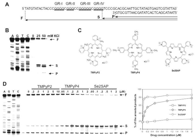
Taq DNA polymerase stop assay showing the stabilization of G-quadruplex structures by K+ and TMPyP2, TMPyP4, and Se2SAP. (A) Sequence of the single-stranded DNA template annealed with primer used in DNA polymerase stop assay. (B) KCl-dependent pausing of DNA polymerase DNA synthesis at the 3′ side of the fourth guanine run in the DNA template containing the VEGF-Pu20 sequence. (C) Structures of TMPyP2, TMPyP4, and Se2SAP. (D) Stabilization of G-quadruplex structure formed by the VEGF-Pu20 with the addition of increasing concentrations of TMPyP2, TMPyP4, and Se2SAP (left panel). Arrows indicate the positions of the full-length product (“F”) of DNA synthesis, the G-quadruplex pause sites (“S”), and the free primer (“P”). Lanes A, G, T, and C represent dideoxysequencing reactions with the same template as a size marker for the precise arrest sites. The percentage of the major arrest products of each sample to the total product was plotted against drug concentrations (right panel)