
Fig. 8.
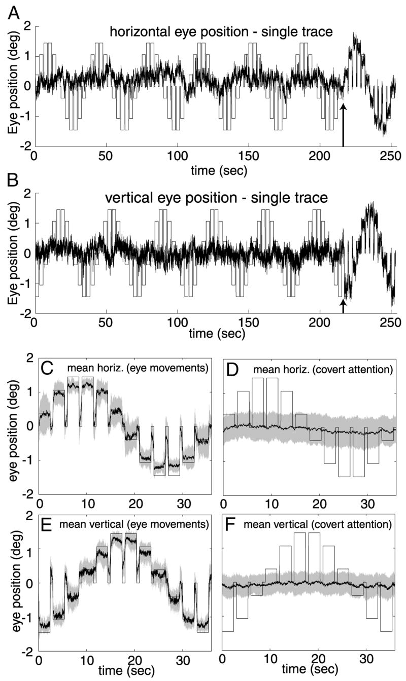
Eye position during attention mapping. A: single example of horizontal eye position recorded in the MR scanner from subject DJH. Black trace, measured eye position. Bars, expected horizontal eye position if the subject made eye movements to the attended segment on every trial. At the time point indicated by the arrow, the screen was blanked for 1.2 s, instructing the subject to make saccades to the attended segment on the subsequent trials. B: vertical eye position from the same example shown in A. C: mean horizontal eye position during performance of the eye movement trials for subject DJH. Shaded region, ±1 SD. D: mean horizontal eye position during performance of the covert attention task. E: mean vertical eye position during eye movement trials. F: mean vertical eye position during covert attention trials. Eye position was very weakly correlated with the cued location when the subject was instructed to maintain fixation.