
Fig. 2. Electrically-aligned astrocytes guide neurite outgrowth.
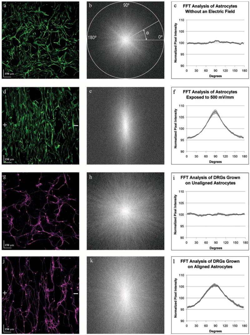
Astrocyte cultures were either exposed to an electric field (500 mV mm−1) for 24 hours or left unexposed as controls. A suspension of dorsal root ganglia cells was then seeded onto the astrocyte cultures and allowed to grow for 48 hours without an electrical field. Images derived from the same field of view show the spatial relationship between control astrocytes (a) and dorsal root ganglion cells (g) and between exposed astrocytes (d) and the dorsal root ganglion cells (j). Plus (+) and minus(−) signs indicate the direction of the electric field vector. (a) Control astrocytes expressing the intermediate filament Vimentin (green), appeared to be randomly oriented. FFT analysis of 12 images provided information about gross image orientation in the form of pixel intensity (b, e, h, k). The pixel intensity of the FFT image is summed along a straight line radiating at the angle, from the center to the edge of the image. (c) Plotting the normalized summed pixel intensity shows equal orientation in every direction. (d) On exposure to an electric field astrocytes display a strong alignment perpendicular to the field. (e,f) FFT analysis of six astrocyte images reveals strong orientation towards 88.8°. (g–i) Similarly, dorsal root ganglia cells seeded on control astrocytes (g) show random orientation that is identical to the astrocyte cultures (h,i). (j–l) Conversely, DRGs seeded on electrically aligned astrocytes (j) preferentially align towards 88.8° (k,l).