
Fig. 3. Neurites follow the processes of electrically-aligned astrocytes.
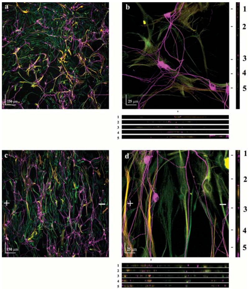
Astrocyte cultures were either exposed to a electric field (500 mV mm−1) for 24 hours or left unexposed as a control. A dorsal root ganglia cell suspension was then seeded onto the astrocyte cultures and allowed to grow for 48 hours. (a,c) Cocultures of neurons (TUJ1+, magenta) and astrocytes (Vimentin+, green and GFAP+, red, which are yellow when colocalized) of control and electrically aligned cultures, respectively. + and − indicate the direction of the electric-field vector. (b,d) Higher magnification indicates that direct contact between neuronal processes and astrocytes is established. Five XZ sections (location marked 1–5) show direct contact between neurites and astrocyte processes in all three dimensions.