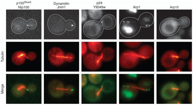
Figure 1. Localization of dynactin in cells.
Fluorescent-tagged forms of dynactin subunits are observed at plus ends of microtubules and at SPBs. The α-tubulin subunit Tub1 was tagged with CFP for colocalization in each strain. The width of each image is 5 μm. Strain numbers: yJC3891, Yll049w-3GFP; yJC4147, Nip100-3GFP; yJC5261, Jnm1-3GFP; yJC5389, Arp10-3GFP and yJC5400, Arp1-tdimer2.

